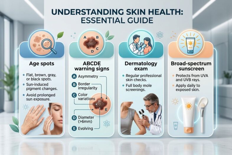
You know how easy it is to dismiss a flat brown mark as “just an age spot”, especially when it appears on the hands or face after years of sun exposure. The key distinction is that age spots are usually harmless, but a changing spot should never be self-diagnosed.
Also called liver spots or solar lentigines, these marks develop when ultraviolet (UV) light triggers extra melanin production in the skin. They are a visible sign of cumulative UV damage, which is why they deserve a closer look rather than a quick cover-up.
This guide explains what age spots look like, how to spot possible skin cancer or melanoma, when to book a dermatologist appointment, and which treatments and prevention steps make sense.
Key Takeaways
- Age spots are flat tan, brown, or dark brown marks caused by long-term ultraviolet (UV) exposure and excess melanin in the skin.
- Typical solar lentigines are benign, but a new, changing, itchy, bleeding, crusted, or uneven spot needs a dermatologist assessment.
- Use the ABCDE warning signs for melanoma: asymmetry, border, colour, diameter, and evolving changes.
- Only treat a spot after a medical dermatology exam confirms that it is benign. A skin biopsy may be needed if the diagnosis is unclear.
- Daily broad-spectrum sunscreen, protective clothing, shade, and avoiding tanning beds help prevent further UV damage and new age spots.
What Are Age Spots?
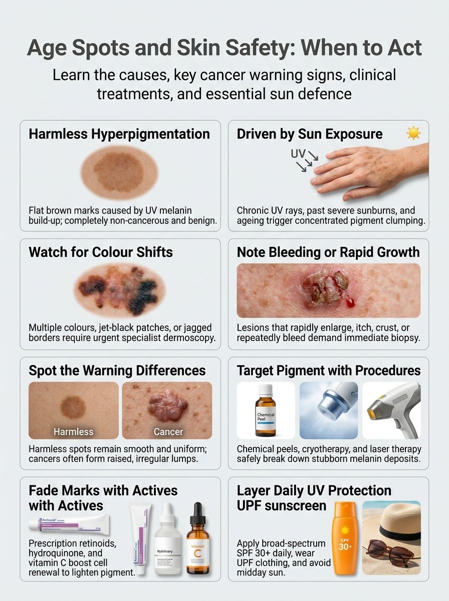
Age spots are small, flat areas of hyperpigmentation. You may also hear them called liver spots, sunspots, or solar lentigines.
They develop when pigment cells produce and deposit extra melanin after repeated sun exposure. Their colour can range from light tan to dark brown, and they often appear on the face, backs of the hands, shoulders, chest, forearms, and tops of the feet.
Most age spots measure a few millimetres across, although larger spots can reach about 13 millimetres. They usually have a smooth surface and sit flat against the surrounding skin.
Age spots become more common with age because UV damage builds over time. The National Institute on Aging describes age spots as harmless, but that does not mean every brown mark is automatically safe to ignore.
- Typical solar lentigo: flat, evenly coloured, and slow to change.
- Possible look-alike: melanoma, lentigo maligna, seborrhoeic keratosis, actinic keratosis, or post-inflammatory hyperpigmentation.
- Best next step: arrange a dermatologist review if one spot looks noticeably different from the rest.
Causes of Age Spots
Long-term sun exposure is the main cause of age spots. Ultraviolet radiation damages skin cells and signals pigment cells to make more melanin, which can collect unevenly and create visible brown patches.
Both UVA and UVB rays matter. UVA reaches deeper into the skin and contributes to premature ageing and pigment changes, while UVB is a major cause of sunburn.

- Repeated outdoor exposure: Driving, gardening, walking, sport, and outdoor work can add up over the years, especially on the face and hands.
- Sunburn: A history of blistering or repeated sunburn adds to UV damage and can make uneven pigmentation more likely.
- Tanning beds: Indoor tanning exposes skin to intense UV radiation. The CDC advises avoiding tanning beds and sunlamps because they increase skin cancer risk.
- Age: Age spots often become more visible after age 40, although significant sun exposure can bring them on earlier.
- Inflammation or injury: Acne, eczema, burns, and skin irritation can leave dark marks called post-inflammatory hyperpigmentation, which may resemble sunspots.
One practical point matters here: a brown mark caused by inflammation may need a different treatment from a true solar lentigo. That is why an in-person diagnosis should come before you use hydroquinone, retinoids, chemical peels, or laser treatments.
Are Age Spots Dangerous?
Age spots themselves are not skin cancer. A confirmed solar lentigo does not turn into melanoma, basal cell carcinoma, or squamous cell skin cancer.
Still, age spots show that your skin has received enough ultraviolet (UV) light to cause pigment damage. That same UV exposure raises the risk of skin cancer, so the real concern is not the age spot alone. It is the possibility that a suspicious lesion is being mistaken for one.

A brown spot that changes in size, shape, colour, texture, or symptoms deserves a professional skin check.
In its 2026 estimates, the American Cancer Society projects about 112,000 new melanoma diagnoses in the United States. Melanoma accounts for a small share of skin cancer cases, yet it causes a large share of skin cancer deaths, which is why early assessment matters.
See a dermatologist in North Central Phoenix, Arizona, or near you if you notice a spot that is new, growing, bleeding, crusting, painful, itchy, or unlike your other marks. A dermatologist can use dermoscopy, close-up skin photography, and a skin biopsy to tell whether a lesion is benign.
When to See a Dermatologist
You should book a dermatologist appointment when a mark changes or simply does not fit the pattern of your other age spots. A quick examination can often provide reassurance, while a suspicious lesion may need a biopsy.
Do not try to fade a questionable spot with over-the-counter acids, retinoids, or bleaching creams before it has been examined. The American Academy of Dermatology warns that treating what appears to be an age spot can delay diagnosis if the mark is actually skin cancer.
New or changing spots
A spot does not need to look dramatic to warrant a skin check. Changes over weeks or months are often more meaningful than a single photograph.
- New mark after age 30: have a new mole or pigmented lesion examined, especially if it appears suddenly.
- Growth: book an appointment if a spot becomes wider, thicker, raised, or develops a new uneven shape.
- Surface change: persistent scaling, crusting, bleeding, or a sore that does not heal needs prompt review.
- Symptoms: itching, tenderness, pain, or recurring irritation can be warning signs.
- Tracking: take clear photographs in the same light and include a ruler or coin for scale, then bring those images to your appointment.
Dermatoscopy, also called dermoscopy, allows a dermatologist to examine pigment patterns below the skin surface. If the lesion still looks uncertain, a skin biopsy lets a pathologist check the tissue under a microscope.
Signs of skin cancer to watch for
The ABCDE rule is a useful screening tool for melanoma. It is not a home diagnosis, but it gives you a clear reason to seek medical dermatology care.
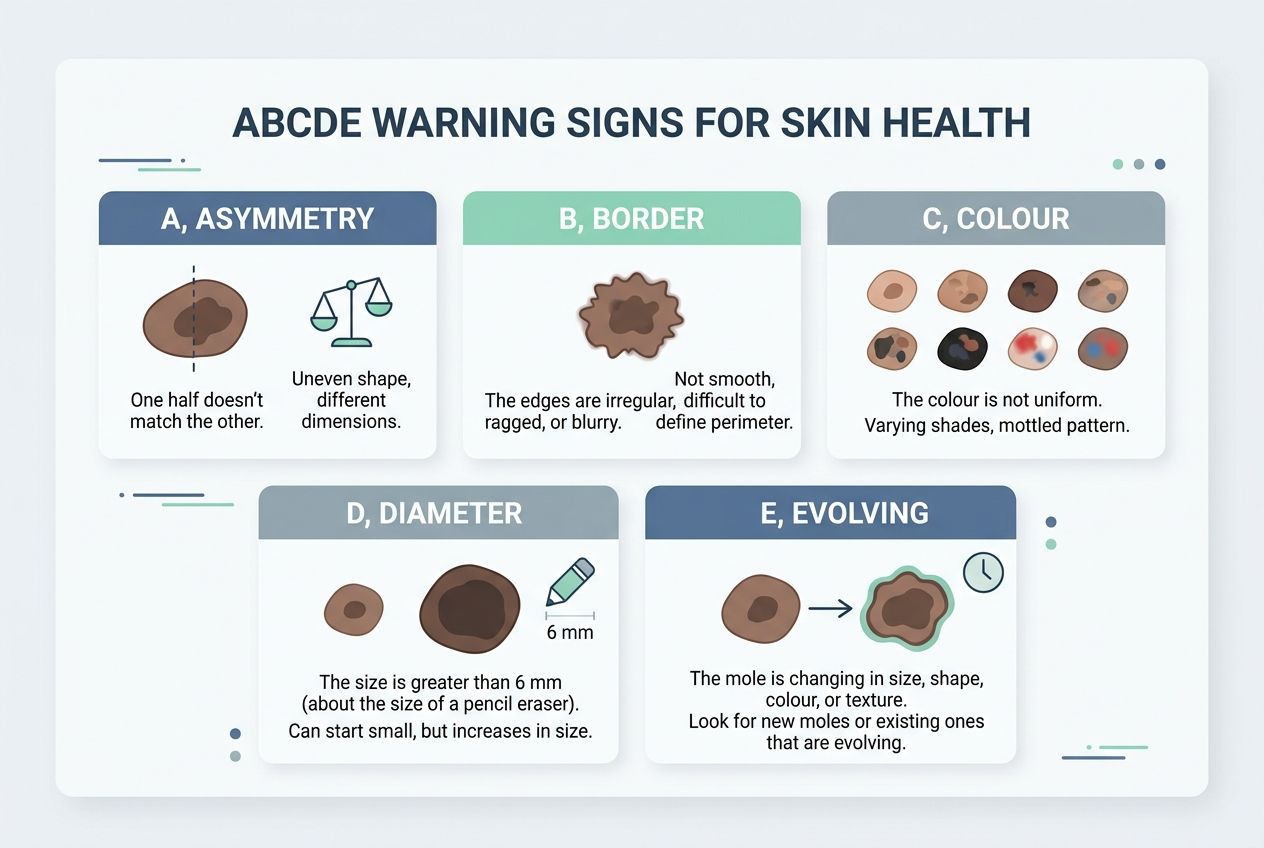
| Warning sign | What to look for | What to do |
|---|---|---|
| A, Asymmetry | One half of the spot does not match the other. | Book a skin check. |
| B, Border | Edges look irregular, scalloped, jagged, or blurred. | Ask for a dermatologist assessment. |
| C, Colour | More than one colour appears, such as brown, black, red, white, blue, or grey. | Arrange a prompt review. |
| D, Diameter | The lesion is larger than about 6 millimetres, although melanomas can be smaller. | Compare it with earlier photographs and seek advice. |
| E, Evolving | The mark changes in size, shape, colour, surface, or symptoms. | Contact a dermatologist without waiting for a routine check. |
Also watch for the “ugly duckling” sign. If one spot looks clearly different from your other freckles, moles, or age spots, it is worth checking even if it does not meet every ABCDE feature.
How to Differentiate Age Spots from Skin Cancer
Age spots and skin cancer can overlap in colour and location, especially on chronically sun-exposed skin. This comparison can help you decide whether you need an appointment, but only a dermatologist can confirm the diagnosis.
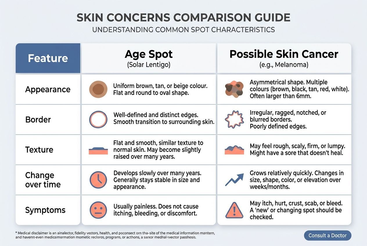
| Feature | Age Spot, Solar Lentigo | Possible Skin Cancer |
|---|---|---|
| Appearance | Flat, tan or brown mark with fairly even colour. | May be dark, mixed in colour, pearly, pink, red, scaly, or unusually pigmented. |
| Border | Usually smooth and well-defined. | May look irregular, blurred, notched, or uneven. |
| Texture | Usually feels like the surrounding skin. | May feel rough, raised, firm, crusted, waxy, or ulcerated. |
| Change over time | Develops slowly and often stays stable. | May grow, bleed, itch, hurt, thicken, or change over weeks or months. |
| Symptoms | Usually painless and symptom-free. | May itch, bleed, crust, sting, or fail to heal. |
| Common sites | Face, hands, chest, shoulders, and forearms. | Can occur anywhere, including the scalp, ears, nails, soles, and areas with little sun exposure. |
| Professional assessment | Visual exam may be enough if the spot is classic and stable. | Dermoscopy, photographic monitoring, and a skin biopsy may be needed. |
Basal cell carcinoma can appear as a pearly bump, a pink patch, or a sore that repeatedly heals and returns. Squamous cell skin cancers may look rough, scaly, crusted, or tender. Melanoma may be flat or raised and can appear in many colours, including skin-coloured or pink.
People with darker skin can develop skin cancer too, including on the palms, soles, under nails, and inside the mouth. Check every part of your skin rather than focusing only on sun-exposed areas.
Treatment Options for Age Spots
Once a dermatologist confirms that a mark is a benign age spot, treatment can fade or remove it. The right option depends on your skin tone, the number of spots, their depth, the body area, and your risk of post-inflammatory hyperpigmentation.
A good treatment plan also includes daily sunscreen. Without UV protection, new age spots can form and treated areas can darken again.
Topical treatments
Prescription treatment may include hydroquinone, tretinoin, or a combination formula chosen by your dermatologist. Hydroquinone reduces melanin production, while retinoids such as tretinoin speed skin-cell turnover.
These products work gradually. Mayo Clinic notes that prescription bleaching creams may fade age spots over several months, so expect steady improvement rather than a change overnight.
- Hydroquinone: can lighten excess pigment, but may cause dryness, redness, or irritation.
- Retinoids: can improve cell turnover and support a more even skin tone, though they may initially cause peeling.
- Vitamin C and niacinamide: over-the-counter options that may help with mild hyperpigmentation when used consistently.
- Glycolic acid or kojic acid: exfoliating ingredients that can irritate sensitive skin if overused.
Apply treatment exactly as directed, and stop if you develop significant burning, swelling, blistering, or a darkening patch. Using a strong active ingredient on an unconfirmed lesion is a common mistake.
Dermatologist-administered procedures
Procedures can provide faster improvement for individual solar lentigines, but they also carry a risk of irritation or pigment changes. This is especially relevant for deeper skin tones and for people prone to dark marks after inflammation.
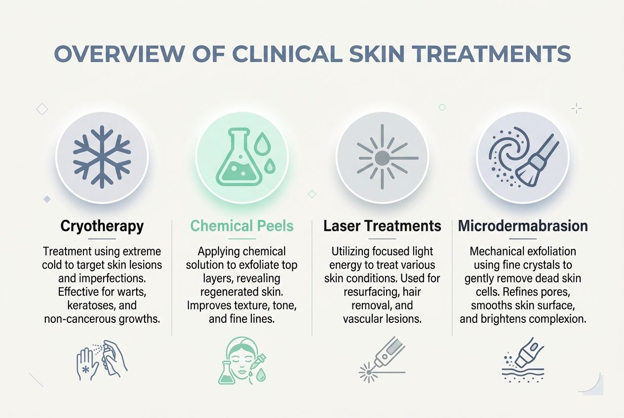
| Treatment | Best suited for | What to expect |
|---|---|---|
| Cryotherapy | One or a few clearly defined age spots. | Liquid nitrogen freezes excess pigment. Blistering, scabbing, and temporary light or dark marks can occur. |
| Chemical peels | Diffuse sun damage or multiple superficial spots. | A controlled exfoliating solution lifts damaged surface cells. Recovery depends on peel depth. |
| Laser treatments or IPL | Multiple brown spots and broader pigment concerns. | Light energy targets pigment. More than one session may be needed, with temporary redness or swelling possible. |
| Microdermabrasion | Mild surface unevenness and gradual brightening. | Mechanical exfoliation with modest results over repeated sessions. |
Microneedling may improve overall texture and some types of hyperpigmentation, but it is not the first choice for every solar lentigo. Your dermatologist should first rule out melanoma and other skin cancers before using any procedure that disrupts the skin surface.
Preventing Age Spots
The most reliable way to prevent new age spots is to reduce UV damage every day. You do not need perfect sun avoidance, but you do need consistent protection.
- Use broad-spectrum sunscreen: Choose SPF 30 or higher for daily outdoor time, and apply it generously to exposed skin, including the ears, neck, hands, scalp, and tops of the feet.
- Reapply on schedule: Reapply at least every two hours outdoors, and sooner after swimming, sweating, or towel drying.
- Use enough product: The FDA advises that an average-sized adult needs about one ounce, roughly a shot-glass amount, to cover the body.
- Check the UV Index: Protect your skin when the UV Index is 3 or higher. In much of the United States, UV rays are strongest from 10 a.m. to 4 p.m. during daylight saving time.
- Wear UPF clothing: Choose long sleeves, trousers, wide-brimmed hats, and ultraviolet protection factor (UPF) clothing where practical.
- Skip tanning beds: A tan is a sign of UV injury, not healthy skin. Tanning beds add concentrated UV radiation and do not create a safe “base tan”.
Sunscreen works best as one layer of protection, not the whole plan. Combine it with shade, clothing, sunglasses, and a hat whenever you expect extended sun exposure.
Risk Factors That May Require Medical Evaluation
Anyone can develop age spots or skin cancer, but some factors make a professional skin check more important. Your personal history matters as much as the appearance of one mark.

- Frequent sun exposure, outdoor work, or repeated sunburns.
- Past or current tanning bed use.
- Fair skin that burns or freckles easily.
- A personal history of basal cell carcinoma, squamous cell skin cancers, or melanoma.
- A family history of melanoma.
- More than 50 moles, atypical moles, or many changing pigmented spots.
- A weakened immune system, including after an organ transplant or during certain medical treatments.
- A new or changing lesion after age 30.
According to the American Cancer Society's 2026 cancer figures, melanoma rates continue to rise slightly among adults aged 50 and older. That makes routine self-checks and prompt evaluation of changes especially useful as you age.
Healthy eating and hydration support general skin health, but they cannot reverse UV damage or replace sunscreen. Vitamin C, vitamin E, green tea extract, and niacinamide may support a skincare routine, yet they should never delay a dermatologist visit for a suspicious spot.
Conclusion
Age spots are common melanin changes linked to ultraviolet (UV) light and years of sun exposure. Most solar lentigines are harmless, but they can look similar to skin cancer.
If an age spot grows, changes colour, bleeds, crusts, itches, or looks unlike your other marks, see a dermatologist for an examination and possible skin biopsy.
Use broad-spectrum sunscreen, cover exposed skin, avoid tanning beds, and treat confirmed age spots only after a medical assessment.
FAQs
1. What are age spots and are they dangerous?
Age spots are flat, brown patches that come from sun exposure and ageing. They are usually not dangerous, but a small number can be skin cancer, so watch them and ask a dermatologist if you are unsure.
2. When should I see a dermatologist about an age spot?
See a dermatologist for a medical check if a spot grows, changes in size or colour, bleeds, itches, or looks different from your other spots. Early checks catch problems sooner, and that lowers risk.
3. How will a dermatologist check an age spot?
The doctor will look closely, often with a special lamp or magnifier, and will note borders, colour and size. If a spot looks odd, they may take a small skin sample, called a biopsy, for lab tests.
4. Can I treat age spots at home, or do I need a dermatologist?
Some creams and over-the-counter products can lighten age spots, but results vary and may take time. For faster, safer results or if a spot worries you, a dermatologist can offer medical treatments such as prescription creams, freezing, laser or removal.